REVIEW
Neuro-ERG: more can be seen
Objective eye examination and analysis of the functional activity of the retina
The Neuro-ERG electroretinograph allows you to perform the following types of examinations:
- rod electroretinography (Dark-adapted 0.01 ERG);
- maximum electroretinogram (Dark-adapted 3.0 ERG);
- cone electroretinography (Light-adapted 3.0 ERG);
- electroretinography pattern;
- Local electroretinography (Macular or Focal ERG)
- rhythmic electroretinography (Light-adapted 3.0 Flicker ERG);
- on/off electroretinogram (Long-duration Light-adapted ERG);
- Oscillatory potentials (Dark-adapted 3.0 Oscillatory Potentials ERG);
- visual evoked potentials (VEP) of the brain per flash of light and reversed pattern;
- rhythmic visual evoked potentials;
- electrooculography (EOG);
- multifocal electroretinography;
- multifocal visual evoked potentials;
- objective assessment of visual acuity.
Exam
nation with the Neuro-ERG electroretinograph can be carried out in both adults and children from the first days of life. Neurosoft owns unique methodological developments in the study of evoked brain potentials in newborns and young children (patent RF No2158534 dated 10.11.2000).
Neuro-ERG complies with the standards approved by the International Society for Clinical Electrophysiology of Vision (ISCEV).
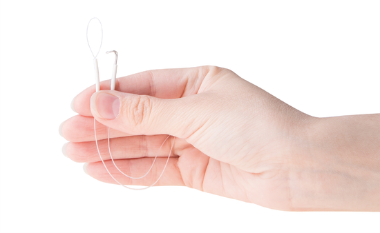
A set of specially designed electrodes ("hooks" and "loops")
The device is accompanied by a set of unique ERG electrodes, specially developed with the participation of Russia's leading specialist in the electrophysiology of vision, Chief Researcher of the Helmholtz Moscow Research Institute of Eye Diseases, Doctor of Medical Sciences A.M. Shamshinova.
The electrodes are designed in the form of small "hooks" and "loops". This type of electrodes does not cause discomfort to the patient during the examination and is very convenient for the doctor when working. In clinics, "suction lenses" electrodes are often used, which prevent the patient from concentrating on the examination due to their "excessive mobility". For the same reason, there is a lot of interference, which affects the quality of the signal.
Moreover, experts consider the use of "suction lenses" to be incorrect in some examinations (for example, with local or cone ERG), since the most important condition is violated: the samples must be carried out under photopic conditions.
Mini ganzfeld stimulator for basic tests
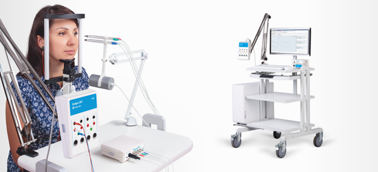
For the main tests (cone, rod, rhythmic ERG, oscillator potentials), a mini-ganzfeld stimulator developed by Neurosoft is designed to have no analogues in Russia, which fully complies with ISCEV standards in terms of its characteristics.
The compact design of the stimulator provides convenience for the patient during the examination and obtaining high results.
Stimulator "light pencils" with concentrators for local tests
To perform such tests as local or cone ERG for color stimuli, a stimulator "light pencils" with concentrators (red, blue, green, white) is used. It is mounted on a special tripod, which solves the problem of fixing the stimulator near the pupil.
Solving a number of clinical problems
ERG allows you to determine the localization of the pathological process in the outer and inner layers of the retina, in its central and peripheral zones. The method makes it possible to study the activity of the rod and cone systems separately.
An important aspect of the method is the diagnosis of initial preclinical changes in the retina. Changes in the electroretinogram are characteristic of many retinal diseases and allow you to assess the degree of damage.
ERG is also necessary for the differential diagnosis of diseases of the retina and optic nerve. In this case, it is carried out together with the study of visual evoked potentials of the brain. Electroretinography, like other neurophysiological research methods, is nosological nonspecific. By the type of ERG, it is impossible to determine which factor caused its change. Despite this, the correct choice of conditions for registering ERG allows you to solve a wide range of diagnostic problems, which makes the study indispensable in the daily practice of an ophthalmologist.
The electroretinograph is included in the standard equipment of the ophthalmology department and day hospital in accordance with the Order of the Ministry of Health of the Russian Federation dated November 12, 2012 No 902n (Appendix No 14) "Procedure for providing medical care to the adult population with diseases of the eye, its appendages and orbit".


